The plant on your windowsill is buzzing with life, turning
sunlight into sugar all day long. Mold is slowly gobbling up the
apples in your fruit bowl. Your bed is creeping with dust mites. The
air is packed with pollen...
It's a truly amazing thought: there are
zillions of things happening all around us, all the time, that are
far too tiny for our eyes to see! But never fear, because we have an
equally amazing way to get around it. Powerful microscopes shed
new light on the teeny tiny and make the invisible, visible. They've
played an enormous part in science by taking us deep into worlds
we've come to think of as "microscopic." Just as telescopes scale
us up to meet the planets and stars, so microscopes scale us down
into the tiny world of atoms and cells. Let's
take a closer look at how they work!
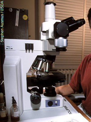
Photo: This looks like an ordinary optical microscope, but it's
actually a fluorescence microscope (a Nikon Eclipse) designed to produce higher-contrast images.
Exactly how it works is described below. Photo by Stephen Ausmus courtesy of US Department of Agriculture: Agricultural Research Service (USDA-ARS).
Lots of things are invisible, but that doesn't mean they're not
there. Radio and TV broadcasts are constantly whistling through your
head from powerful transmitters, but unless you happen to have a
cunning piece of electronic equipment at your disposal—namely a
radio or TV
set—you won't be able to understand them. We're used to
the world being the totality of things we can see; that there are
worlds out there our eyes aren't tuned into is both a physical
problem and a philosophical conundrum.
Imagine if your eyes were as powerful as microscopes and you could
see all the germs crawling about on your hands. Your brain would be
so busy boggling that you wouldn't be able to concentrate on bigger
things at a more meaningful scale. Through millions of years of
evolution, our eyes and brains are programmed to worry about the
things that matter most—things on a similar scale to
our bodies. We simply don't have the time or the brain capacity to
worry about absolutely everything that's going on. If you were
constantly staring at the bugs on your fingers, you could easily get
so distracted that you'd walk straight under a bus!
Don't understand? Let's put it this way. The smaller
the things you look at, the more there is to see, the more information
there is to process, and the longer it takes. If you could see
microscopically all day long, you'd have to react much more slowly
to the world around you—and that extra reaction time would threaten your life.
This, then, is what invisible means: our bodies are finely tuned to the
business of day-to-day living on a human scale and efficiently designed to ignore
everything else.
Once upon a time, we used to ignore things we couldn't see. But
thanks to modern science, we know there's a whole lot happening on
the microscopic scale that can help us to live our lives more
effectively. Scientists have known since the 17th century that the
insides of living things are made up of tiny functioning factories
called cells; understanding how they work helps us to tackle sickness
and disease. More recently, during the 20th century, scientists
figured out how materials are made of atoms and how atoms themselves
are built from smaller "subatomic" particles; understanding
atomic structure paved the way for all kinds of amazing inventions,
from electronic transistors to nuclear power.
Sponsored links
How microscopes work
Photo: Most microscopes have several different objective lenses that turn around on a thumb-wheel to give different levels of magnification. Going from right to left, the lenses you can see here magnify by twenty times (20x), forty times (40x), and a hundred times (100x). Photo by Stephen Ausmus courtesy of
US Department of Agriculture: Agricultural Research Service (USDA-ARS).
Microscopes are effectively just tubes packed with lenses, curved
pieces of glass that
bend (or refract) light rays passing through them.
The simplest microscope of all is a magnifying glass made from a single convex lens, which
typically magnifies by about 5–10 times. Microscopes used in homes, schools, and professional laboratories are actually compound microscopes and use at least two lenses to produce a magnified image. There's a lens above the object (called the objective lens) and another lens near your eye (called the eyepiece or ocular lens). Each of these may, in fact, be made up of a series of different lenses.
Most compound microscopes can magnify by 10, 20, 40, or 100 times, though professional ones can
magnify by 1000 times or more. For greater magnification than this, scientists generally use
electron microscopes.
Photo: Ordinary microscopes are "powered" by light. When light shines on the specimen at the bottom, it travels straight through or reflects off the surface, passing up through the lenses into the eyepiece. Microscopes that use light are called optical microscopes to distinguish them from electron microscopes, which use electrons for seeing instead of light. Photo by Peggy Greb courtesy of US Department of Agriculture: Agricultural Research Service (USDA-ARS).
So what does a microscope actually do? Imagine a fly sitting on the table in front of you. The big, fat, compound eye on the front of its head is just a few millimeters across, but it's made up of around
6000 tiny segments, each one a tiny, functioning eye in miniature. To
see a fly's eye in detail, our own eyes would need to be able to process
details that are millimeters divided into thousands—millionths of a
meter (or microns, as they're usually called). Your eyes may be good,
but they're not that good. To study a fly's eye really well, you'd
need it to be maybe 10–100 cm (4–40 in) across: the sort of size
it would be in a nice big photo. That's the job a
microscope does. Using very precisely made glass lenses, it takes the
minutely separated light rays coming from something tiny (like a fly's eye) and spreads
them apart so they appear to be coming
from a much bigger object.
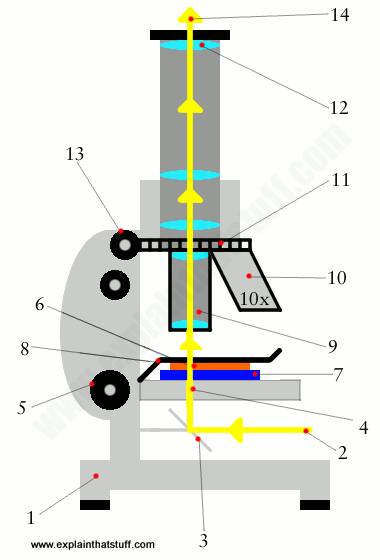
Parts of a microscope
A compound microscope uses two or more lenses to produce a magnified image of an object, known as a specimen, placed on a slide (a piece of glass) at the base.
The microscope rests securely on a stand on a table.
Daylight from the room (or from a bright lamp) shines in at the bottom.
The light rays hit an angled mirror and change direction, traveling straight up toward the specimen. The mirror pivots. You can adjust it to capture more light and alter the brightness of the image you see.
The light rays pass through a hole in an adjustable horizontal platform called the stage.
The stage moves up and down when you turn a thumb wheel on the side of the microscope. By raising and lowering the stage, you move the lenses closer to or further away from the object you're examining, adjusting the focus of the image you see.
To look at something under a microscope (such as a plant leaf), you prepare a specimen of it. The specimen has to be a very thin slice so light rays will pass through.
You mount the specimen on a glass slide with a glass cover slip on top to keep it in place.
The slide is held in place by two metal clips, one on either side.
Light traveling up from the mirror passes through the glass slide,
specimen, and cover slip to the objective lens (the one closest to the object). This makes the first magnification: it works by spreading out light rays from the specimen so they appear to come from a bigger object. The objective "lens" usually consists of more than one lens.
A selection of other objective lenses can be used to magnify the specimen by more or less.
The thumb wheel makes it easy to swing the other lenses into position.
The eyepiece lens (the one closest to your eye) magnifies the image from the objective lens, rather like a magnifying glass.
On some microscopes, you can move the eyepiece up and down by turning a wheel. This gives you fine control or "fine tuning" of the focus.
You look down on a magnified image of the object.
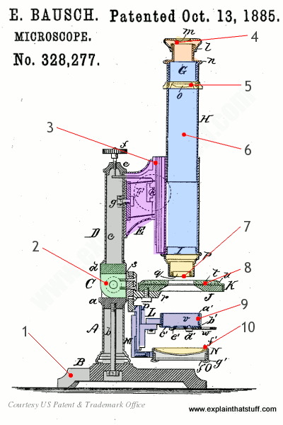
Artwork: A classic optical microscope from the late 19th century.
From US Patent 328,277: Microscope by Edward Bausch, granted October, 13, 1885, courtesy of US Patent and Trademark Office.
That's the theory; here's what it looks like in practice. This is a diagram of a classic Bausch microscope from 1885, and you can see straight away that little has changed over the last century or so. I've colored the main parts and simplified the numbering to make it easier to understand. Briefly, we have:
A heavy metal base gives the microscope a low center of gravity so it's less likely to tip over. The rest of the microscope is made of lightweight sheet metal to keep the cost and weight down.
A hinge allows the main optical tube to pivot up and down.
The focusing mechanism on this scope uses a rack and pinion gear to bring the lenses nearer to or further from the object you're viewing.
The eyepiece (orange) and eyepiece lens (yellow).
A field lens (yellow) ensures more of the light from the object goes into the eyepiece.
The main imaging tube is made from spun or stamped sheet metal.
The objective lens.
The stage and glass slide.
The sub-stage includes an iris (diaphragm) to regulate the amount of light passing through.
One or more mirrors for catching and reflecting light up through the slide.
Polarizing microscopes
As we've just seen, a basic optical microscope uses one or more lenses to bend the light bouncing off (or traveling through) a
specimen, so fooling our brains into thinking what we're looking at is bigger than it really is. But there are other kinds of optical microscopes that work in different ways.
Ordinary light consists of waves that vibrate in every direction. If we pass light like this through a grid-like
filter, so the waves can vibrate in only one direction (plane), what we get is called polarized light (or, sometimes,
plane-polarized light). If you shine polarized light through an ordinary piece of glass, it bends in the usual way
through the process of refraction (or refringence, as it was once known). We can compare the amount by which light bends in different materials using a measurement called the refractive index. (Refraction is explained in more detail in our main article about light.)
So far so good. But other solid materials, including ice, calcium carbonate, quartz, plastics (such as cellophane),
stressed plastics, and various other crystals, change light in different ways when it travels through them in different directions. If you pass light through these materials, something much more interesting happens: it splits into two separate waves that bend by different amounts. In other words, unlike glass (which has a single refractive index), these materials have two refractive indices; that's why this effect is called birefringence (in effect, double refraction). If you collect the two waves coming out of a birefringent crystal and pass them through another polarizing filter, called an analyzer, which is set up at right angles to the first polarizing filter, they recombine, interfere with one another, and produce colorful patterns that change as you rotate the specimen.
Photo: Photoleasticity means using polarized light to study the stress patterns in a material.
In this example, I'm making some polarized light using an ordinary LCD laptop
screen, passing it through a plastic CD case, and viewing the result through polarizing sunglasses.
Polarizing microscopes work in a broadly similar way.
Polarizing microscopes are based on this idea: they're much like ordinary optical microscopes but with polarizing filters fitted above and below the specimen. We can use them to study birefringence and other properties of materials, which can help us identify minerals or figure out important things about their inner structure. Polarizing microscopes have useful applications in:
Geology—for example, studying the mineral components of a particular rock.
Crystallography—for identifying crystals in everything from
forensic science to art conservation.
Seeing things under microscopes isn't just a matter of making them look bigger; a lot
of the time, the problem is making things stand out enough so we can see them at all. In other
words, it's a matter of improving the contrast in an image.
Photo: Increasing contrast makes it easier to see an object against its background and identify what it is from its key features ("specificity").
Optical microscopy uses all sorts of
tricks—chemical and physical—for achieving this.
If you've ever used a simple microscope at school, for example, you probably used chemical stains like iodine,
which turns carbohydrates a dark brown or black, or
methylene blue (C16H18N3ClS), which shows up the shape of bacteria by coloring acids inside them a deep shade of blue. As we saw up above, polarizing microscopes achieve colorful contrast with the help of
polarized light waves. And there's another very popular, contrast-improving technique in microscopy that relies
on fluorescence—the chemical trick pulled by creatures like fireflies.
Photo: Looking at a high-contrast image of an Indian meal moth caterpillar
using a fluorescence microscope. Fluorescence from the sample is picked up by an image sensor and displayed on a computer screen. Photo by Greg Allen courtesy of US Department of Agriculture: Agricultural Research Service (USDA-ARS).
In a fluorescence microscope (like the one in the top picture), we stain the specimen with a fluorescent dye,
and bombard it with light of a particular wavelength (usually from a mercury lamp). That makes the dye give off light of a different (longer and redder) wavelength by the process of fluorescence, which produces a high-contrast
image we can look at through conventional lenses (using special filters) or record with a camera (or a light-detecting image sensor).
Fluorescence microscopes are very widely used because they have greater contrast and sensitivity than ordinary
scopes, and they help pick out the unique, functional bits and pieces in microscopic
samples that make it easier to identify things (technically, this is called greater "specificity").
Artwork: How a fluorescence microscope works.
Light from a mercury arc lamp (1) passes through a filter (2) that selects
a particular wavelength we want to excite our sample.
The beam hits a dichroic mirror (3), which reflects certain wavelengths and allows
others to pass through, and bounces down onto our specimen at the bottom (4).
The specimen fluoresces and gives off a redder beam of light (5) that
passes back through the dichroic mirror (6) and a filter into our eyepiece or image sensor (7).
Electron microscopes
Even the best microscopes have their limits: most can't magnify
more than a few thousand times. Why? One way of understanding light is
to say that it's made up of energetic particles called photons,
which behave like waves hundreds of times thinner than a human hair. But what if we
want to look at things smaller than this? In that case, we can't use
light: we have to use particles with even smaller wavelengths—namely electrons,
the tiny negatively charged particles that whizz around inside
atoms. Microscopes that work in this way are called
electron microscopes.
Who invented microscopes?
Here's a brief history of some key moments in microscopy:
c.1595: Dutch spectacle maker Hans Janssen and his son Zacharias develop the first compound microscope.
1665: Robert Hooke publishes Micrographia, showing amazing studies of living things seen and drawn using a microscope.
1675: Dutch businessman Antonie van Leeuwenhoek develops some of the first practical microscopes using high-quality glass lenses and uses them to make the first observations of bacteria and protozoa.
1815: Film photography pioneer William Henry Fox Talbot and (later) physicist David Brewster develop the polarizing microscope.
1873: German physicist Ernst Abbe notes that the fundamental nature of light sets limits on what can be seen ("resolved") with conventional, optical microscopes—the theoretical insight that leads to electron microscopes.
1911: Carl Zeiss and Carl Reichert develop the fluorescence microscope.
1931: German scientists Max Knoll and his pupil Ernst Ruska develop the magnetic electron objective lens, the core component of an electron microscope.
1932: Frits Xernike invents the phase contrast microscope.
1933: Ernst Ruska builds the first practical transmission electron microscope.
1935: Max Knoll builds the first scanning electron microscope (SEM).
1981: Gerd Binnig (1947–) and Heinrich Rohrer (1933–) invent the scanning tunneling microscope (STM), for which (along with Ernst Ruska) they're awarded the 1986 Nobel Prize in Physics.
Microscope history: A great website about historic microscopes and the development of microscopy through the ages, from the Whipple Museum of the History of Science at Cambridge University, which has a collection of over 400 important microscopes.
Microscopes: Help Scientists Explore Hidden Worlds: This nice interactive Nobel Prize website explains four of the newest types of microscope and allows you to compare different objects as they'd appear through them. [Archived via the Wayback Machine]
Molecular Expressions: A very comprehensive website dedicated to all the different kinds of optical microscopy, compiled by the late Michael W. Davidson and his colleagues at Florida State University.
Microscopy-UK: A useful collection of articles and other online resources for microscopy professionals and interested amateurs.
Microscopy Society of America: An organization for amateur enthusiasts and microscopy professionals alike, founded in 1942.
Books
For older readers
Introduction to Optical Microscopy by Jerome Mertz. Cambridge University Press, 2019. A comprehensive guide to all the different kinds of optical microscope, including phase contrast, holographic, and more. (Mertz is a professor of biomedical engineering at Boston University, specializing in microscopy.)
Optical Microscopy by Brian Herman. Academic Press, 1993. Slightly dated now, but still well worth a look.
For younger readers
The Usborne Complete Book of the Microscope by Kirsteen Rogers. Usborne, 2012. A great starting point for microscopy-happy kids (suitable for ages 9+), written with the usual Usborne clarity and user-friendliness.
Adventures with a Microscope by Richard Headstrom. Dover, 2012 (reprint). A 232-page guide containing 59 simple microscope adventures. Originally published in 1941, but the basic concepts don't date. Great for young scientists aged 10+.
The World of the Microscope by Chris Oxlade. Usborne, 2008. Award-winning introduction to microscopes, probably best for older children (ages 10+).
The Ultimate Guide to Your Microscope by Shar Levine and Leslie Johnstone. Sterling, 2008. A comprehensive, 143-page guide for children, ages 10+, that describes how to use microscopes and slides safely, then moves on to a range of themed activities (looking at things like food, water, the human body, and imagined crime scenes).
Microlife That Lives in Soil by Steve Parker. Raintree, 2005. A series of 32-page books by Steve for ages 8–10 that also includes Microlife That Helps Us,
Microlife That Makes Us Ill, and Microlife That Rots Things—with an emphasis on microorganisms and cool micrographs in each case.
Microscopes by Adele Richardson. Capstone, 2004. A short and simple (but still useful) guide, probably best for curious children ages 6–9.
Articles
Popular science
DNA Microscope Sees 'Through the Eyes of the Cell' by Knvul Sheikh. The New York Times, June 20, 2019. A new microscopy technique uses DNA and computer processing to map the locations of molecules inside cells.
Please do NOT copy our articles onto blogs and other websites
Articles from this website are registered at the US Copyright Office. Copying or otherwise using registered works without permission, removing this or other copyright notices, and/or infringing related rights could make you liable to severe civil or criminal penalties.